Menu

Cath Lab Procedure

Cardiovascular Treatments & Services

Our team of cardiovascular specialists offers extensive experience in all aspects of cardiology care: non-invasive cardiology, interventional cardiology, and electrophysiology. Each doctor brings a unique perspective and offers specialized care in the detection and treatment of heart disease, peripheral vascular disease, and disorders of the heart.
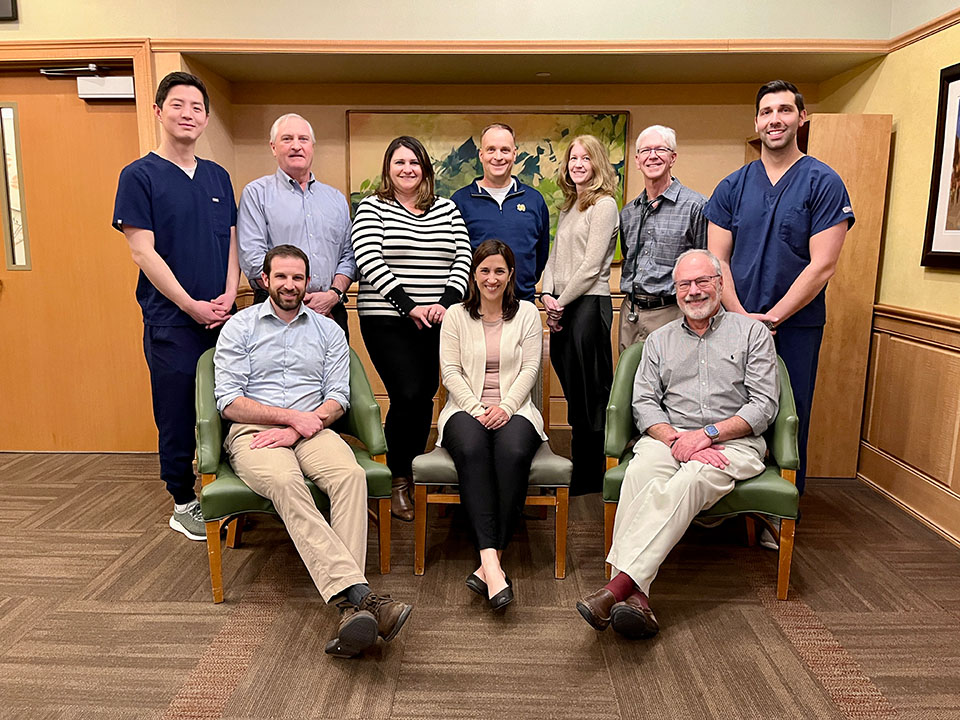
ADVANCED CAPABILITIES

Our team also includes advanced expertise in complete interventional cardiac care, including cardiac catheterization, interventional coronary procedures, and interventional peripheral vascular procedures. We offer office locations in York, Wells, and Newington (formerly known as Seacoast Cardiology) and provide testing and treatment at some of these locations and at York Hospital.


CARDIAC DIAGNOSTIC TESTING AVAILABLE

  •  
Ankle-Brachial (ABI) Testing
A test that uses ultrasound to detect peripheral vascular disease. This test uses doppler in conjunction with a series of arm and leg blood pressure cuffs to measure blood flow in the limbs. This test is most commonly used to determine if leg pain is the result of poor circulation in the legs (claudication). Because these tests use ultrasound and not x-rays, they are completely safe. These procedures do not require any preparation.
Cardiovascular Screening

($100)* This includes screening for abdominal aortic aneurysm (AAA), carotid artery disease, atrial fibrillation and peripheral artery disease (PAD). Rely on the experts at York Hospital to assess your overall cardiovascular health by signing up for one of our low-cost, preventive cardiovascular health screenings! Each 1/2 hour appointment will provide screenings for the most life-threatening vascular diseases. Appointments are necessary. Call York Hospital’s Heart Health Care team at (207) 351-2424 for more information. *These screenings may not be covered by Medicare or other
insurance companies.
Please check with your carrier for details.

Echocardiogram

This test is usually called an Echo. It uses sound waves (ultrasound) to produce a moving, two-dimensional (2D) image of the patient’s beating heart on a video screen. It is a painless procedure that does not use x-rays and is completely safe. The physician can study the heart’s size and function, the motion and structure of the four heart valves and measure the direction and speed of blood flow. The test takes approximately 45 to 60 minutes to perform. No preparation is required.

EKG – Electrocardiogram
An EKG is a graphic display of the electrical activity of the heart from electrodes placed on the skin in specific locations. The electrical activity created by the patient’s heart is processed by the EKG machine and then printed on special graph paper which is later interpreted by a physician. This is a painless process that requires no preparation and takes approximately 10-15 minutes to perform.
Event Recording
It is not uncommon for patients to have symptoms that occur infrequently and are not documented by a 24-hour Holter monitor. This test is similar to a Holter monitor except it is meant to be worn for several days. Instead of the entire 24 hours of EKG information being stored, the event recorder stores or ‘loops’ only the last 40-60 seconds of EKG continuously until a patient has symptoms. The patient then pushes a button on the recorder that ‘freezes’ the current 40-60 seconds stored in memory and the records an additional 15-20 seconds of EKG. The recorder then locks this information in its memory, and goes back to the ‘loop’ mode to wait for the button to be pressed again if there is another event. Typically five events can be saved in the recorder. The event can be transmitted by phone to Cardiology to allow us to look at what has been recorded. The recorder is then reset and ready to record additional events if needed. Patients are shown how to connect themselves to allow them to sleep and bathe. The recorders are usually kept for 1 – 2 weeks.
Holter Monitoring
A continuous, twenty-four hour electrocardiographic (EKG) recording of the heart’s rhythm to determine if rhythm disturbances are present and whether or not they need to be treated. This test is done most commonly for one of three reasons: a patient feels one or more of these disturbances such as palpitations (ectopy) or rapid heart rate (tachycardia); a patient is having symptoms (such as dizziness or fainting) which may be due to tachycardia and/or bradycardia (slow heart rate); To determine if a patient’s pacemaker or defibrillator is working properly. A portable digital recorder about the size of a deck of cards is connected via 5 adhesive electrodes to the patient’s chest. This device records every heartbeat over the 24-hour period it is worn. When it is returned, the information is scanned by special computer software. A diary is given to the patient to document daily activity to allow symptoms and activities to be correlated with the recording.
Stress Echo
A Stress Echo is a non-invasive test that combines two tests, a treadmill test and an echocardiogram. This test is use in situations where it is known that the patient’s EKG will not be able to provide adequate diagnostic information or the physician suspects that a previous stress test was a false positive. The addition of the echocardiogram helps to improve the accuracy of the test by providing additional mechanical information about the heart during exercise. Echo images are taken before and immediately after exercise. These images are then interpreted along with the EKG at the end of the test.
Transesophageal Echocardiogram
A minimally invasive echocardiogram that offers very clear images of the heart’s structures and valves, without the interference of the chest wall and lungs. This procedure requires that a probe be passed down the esophagus (much like a gastroscope), except it uses ultrasound instead of a light source and camera. Total time for the test is about 1 hour. A patient needs to fast after midnight. The time that the probe is in the body is typically about 10 to 15 minutes. An IV is started to administer sedation to make the patient comfortable during the procedure. If the test is performed as an outpatient procedure, the patient needs to arrange for transportation home.
Treadmill Test/Stress Test – Adult
This test evaluates the heart’s response to exercise using a treadmill. It is used primarily to determine if a patient has coronary artery disease. The heart’s electrical activity is continuously monitored using an electrocardiograph (EKG). The treadmill exercise portion of the test is staged. The treadmill starts at a very slow walking pace and at a slight uphill grade. Every three minutes the speed and grade are increased. During the exercise portion of the test, a physician is present. The goal of the test is to achieve an age adjusted target heart rate to make the test statistically reliable. The physician will discuss the results of the test with the patient immediately afterward. The complete procedure takes 30 – 45 minutes, with the exercise portion typically lasting 6-12 minutes. Depending on the reason for the treadmill, some medications should not be taken before the test. It is important to discuss this with your physician prior to the day of the test.
T-wave Aternans Testing
  • This is a specialized type of EKG that looks at a very specific aspect of the heart’s electrical activity. It is used as a screening tool to assess a patient’s risk for a certain type of rhythm abnormality. It is usually done in conjunction with a stress test.
Vascular Ultrasound
This is a painless technique using ultrasound for visualizing arteries and veins. It is a generic term that includes tests such as Carotid Ultrasound, Venous Ultrasound and Arterial Ultrasound. These procedures are sometimes referred to as Duplex Vascular Ultrasound because the information is typically displayed in two different ways. One is an image that displays the cross sectional anatomy of the blood vessel being studied. It is what is typically thought of as an ultrasound image (like the baby pictures of a pregnancy). The other is what is called doppler. This is a two dimensional visual display of the speed and direction of blood flow within a vessel, similar to doppler weather maps that display cloud movements overlaid on a map. Doppler allows us to determine if blood flow has slowed, reversed or stopped in a blood vessel because of disease and accurately measure the severity of the disease.

CARDIAC CATHETERIZATION

York Hospital’s renowned Cardiac & Vascular Catheterization Lab provides cardiac catheterizations, electrophysiology studies, and ablations and vascular & cardiac angiography. In addition, York Hospital is one of only four hospitals in the state of Maine to perform Percutaneous Coronary Interventions (coronary artery ballooning/stenting).

ELECTROPHYSIOLOGY

York Hospitals’ Cardiology team also offers electrophysiology as a specialty. If heart rhythm problems are discovered, physicians and members of the team will perform the appropriate procedures including the implant of a pacemaker (including biventricular) or defibrillator.

PACEMAKER & IMPLANTABLE DEFIBRILLATOR CLINICS

Pacemakers and implantable cardioverter defibrillators (ICD) are small battery-powered, devices implanted in the chest to regulate the heart’s rate and rhythm. Pacemakers are designed to correct bradycardias (slow heart rhythms) and ICD’s tachycardias (fast heart rhythms). These devices are implanted at York Hospital. Patients with pacemakers and ICD’s need to have their devices checked to monitor the clinical effectiveness of their devices and the status of the batteries and electronics. Clinic visits usually occur every three to four months but could be more frequent depending on several factors, such as how old the device is or whether a device needs to be readjusted because of changes in a patient’s medical status. Telephone follow-up is also done for pacemaker patients. Patients use a special transmitter to send their EKG to the clinic by phone. This allows patients the convenience of checking their pacemaker from anywhere they have access to a phone without having to come to the clinic for every checkup. Typically, an individual will be scheduled for telephone pacer checks once every three months, with one clinic visit scheduled per year.

REHABILITATION

Living Well Center: 

Cardiac and Pulmonary Rehabilitation: The Cardiopulmonary Rehabilitation Program is designed to meet the needs of cardiopulmonary patients (including, but not limited to, those who have had a heart attack, coronary artery bypass surgery, Chronic Obstructive Pulmonary Disease, or Angina) and their families. The program combines education, exercise, nutritional counseling, stress reduction, and behavior modification strategies. The goal of Cardiopulmonary Rehabilitation is to help patients recover, resume their normal activities of daily life and develop positive lifestyle strategies to enhance their future health.

We offer specialized clinical exercise and education programs for Cardiac, Pulmonary, Congestive Heart Failure, Diabetes, Oncology, Osteoporosis/Osteopenia, Spine Health and Balance. We also offer many community-based health programs, support groups, disease management education, and more.  Call 207-351-3700 or click this link for more information.